Welcome to the ASBMR Poster Nr. MO0440
You can download the poster in full resolution here: ASBMR2013 Poster Nr. MO0440
SEVERE ALTERATIONS IN CORTICAL AND TRABECULAR BONE MICROARCHITECTURE IN LUNG TRANSPLANT RECIPIENTS

Alexander Valentinitsch, Lukas Fischer, Matthew DiFranco, Claudia Schueller-Weidekamm, Franz Kainberger, Georg Langs, Janina Patsch
Quantitative in-vivo bone imaging techniques including high resolution peripheral quantitative computed tomography (HR-pQCT) have advanced the knowledge on microstructural pathomorphologies of metabolic bone diseases, including postmenopausal and age-related osteoporosis, hyperparathyroidism and diabetic bone disease. Post-processing methods used in cross-sectional and longitudinal HR-pQCT studies have focused on bone geometry, density, and bone microarchitecture, the quantification of cortical porosity, region-specific analyses, and texture-based clustering. Motivated by the latter, we detect differences trabecular bone quality in a female and male cohort of patients with rapid secondary bone loss following lung transplantation (LuTX) in comparison with health controls (Co)
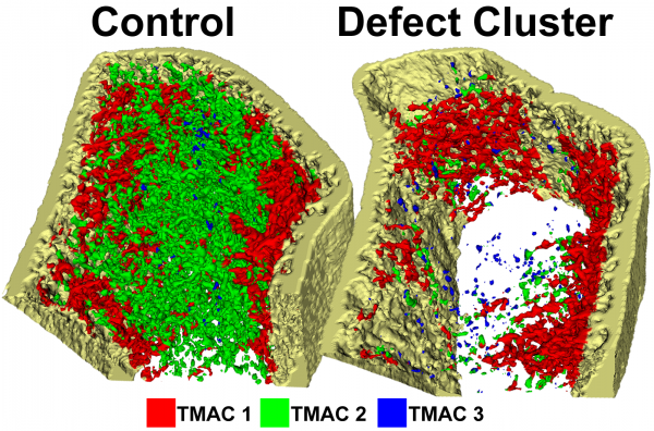
After initial recovery from lung transplantation, 46 patients (female: n=28, male: n=18) were enrolled. Age-matched controls were recrutied (mean age: female 45.8±12.2, male 41.0±15.7). Mean age difference was ±2 years. Scans were analyzed using a 3D texture-based clustering approach for classifying the trabecular compartment into three trabecular microarchitecture classes (TMACs), which are expressed as cluster volume fractions (CL.V/TV) of each class. Standard analysis and cortical porosity analysis from the manufacturers software were also performed. A paired t-test was used to measure statistical significance between LuTX and Co.
The TMAC algorithm differentiates three morphological bone classes (TMAC-1, -2 and -3) that are based on distinct three-dimensional characteristics in trabecular microarchitecture. TMAC-1 predominantly represents thick trabeculae with low intratrabecular spacing. TMAC-3 contains thinner, inhomogeneous trabecular structures that are poorly connected. TMAC-2 represents zones of intermediate morphometric quality.
No significant difference was observed for CL.V/TV of the TMAC-1 (female: p=0.27; male: p=0.53). For the male cohort, a significant difference was observed in TMAC-2 (p=0.001) and TMAC-3 (p=0.02). For females, however, TMAC-2 (p=0.75) and TMAC-3 (p=0.05) did not show a significant difference between the LuTX and Co, likely due to the smaller trabecular region of interest in the female cohort. Cortical porosity parameters were more discriminative in female cohort than in male.
We demonstrated that TMAC algorithm is a promising new HR-pQCT post-processing option.
Longitudinal clinical evaluation of bone architecture and biomechanical changes in transplantation osteoporosis, funded by the Austrian National Bank Anniversary Fund
Team: Lukas Fischer, Rene Donner, Philipp Peloschek, Philippe Zysset, Georg Langs, Janina Patsch
Transplantation osteoporosis is a severe, rapidly progressive type of secondary bone disease. Although early antiosteoporotic treatment can prevent major bone loss, fractures occur. Dual-X ray-absorptiometry (DXA) is unable to monitor small density variations which could be indicative of potential treatment non-responders with elevated fracture risk. Improved risk assessment tools such as FRAX, which are well suitable for classical risk groups such as postmenopausal women, are inapplicable to transplant patients. Moreover, bone metabolism and microarchitectural changes after transplantation are little explored. Using novel imaging tools like high resolution peripheral quantitative computed tomography (HRpQCT), it is nowadays possible to study bone microarchitecture in vivo without biopsy. Thus it is possible to perform detailed, longitudinal monitoring of microarchitectural changes in the individual patient. Moreover, advanced biomechanical evaluations such as computerized compression tests (i.e. finite element modelling) can be derived from such data sets. Advanced computational image analysis is an essential step in the processing of HRpQCT since it offers approaches to efficiently deal with high-dimensional and complex patterns. Using HRpQCT of the distal radius, we will accompany over 100 patients during their first 2 years following lung transplantation (LUTX). Specifically, we aim to establish longitudinal FE modelling and advanced texture-based image analysis as novel, non-invasive monitoring tools for the in vivo-assessment of changes in bone microarchitecture.
