
Emerging Infectious Lung Disease Monitor
ONSET is funded by the Austrian Science Fund (FWF) to develop methods for early detection and management of emerging pandemics.
Team: Georg Langs, Helmut Prosch, Jeanny Pan, Svitlana Pochepnia, Branko Mitic, Konstantin Miloserdov, Lucian Beer, Matthias Perkonigg, Sebastian Röhrich
The project is part of a collaboration network:
AIX-COVNET an international initiative to share data and perform collaborative research to improve diagnosis and prediction for treatment guideance of COVID-19 patients.
ZODIAC Zoonotic Disease Integrated Action of the UNO (IAEA, WHO, FAO)

Project summary
The world-wide spread COVID-19 resulted in a global health care crisis. It made clear that we need the capability to detect an epidemic or pandemic disease early, that we need to be able to diagnose it rapidly, and that we require mechanisms to identify the correct treatment for individual patients. Lung imaging had an important role, shifting from an initially diagnostic- to a prognostic tool informing individual care. The project is a close interdisciplinary collaboration between experts in machine learning and radiology. It will develop new methods in the area of machine learning and image analysis to address these challenges. It will investigate and advance methods for the detection of anomalies and create techniques for the identification of newly emerging phenotypes in the patient population. This will be based on imaging data and clinical information of patients, and is challenging since it involves identifying markers that are not yet known. The second main aim is to develop models that can predict the trajectories of individual patients during their disease and recovery to guide optimal individual treatment. Here, the challenge is to learn from the real-world data collected during the early phase of a pandemic, when no treatment guidelines are available, and the observed patient histories are very diverse. The project will be embedded in international collaborations to ensure the validation of the novel methodology.
Predicting severity of disease based on imaging data
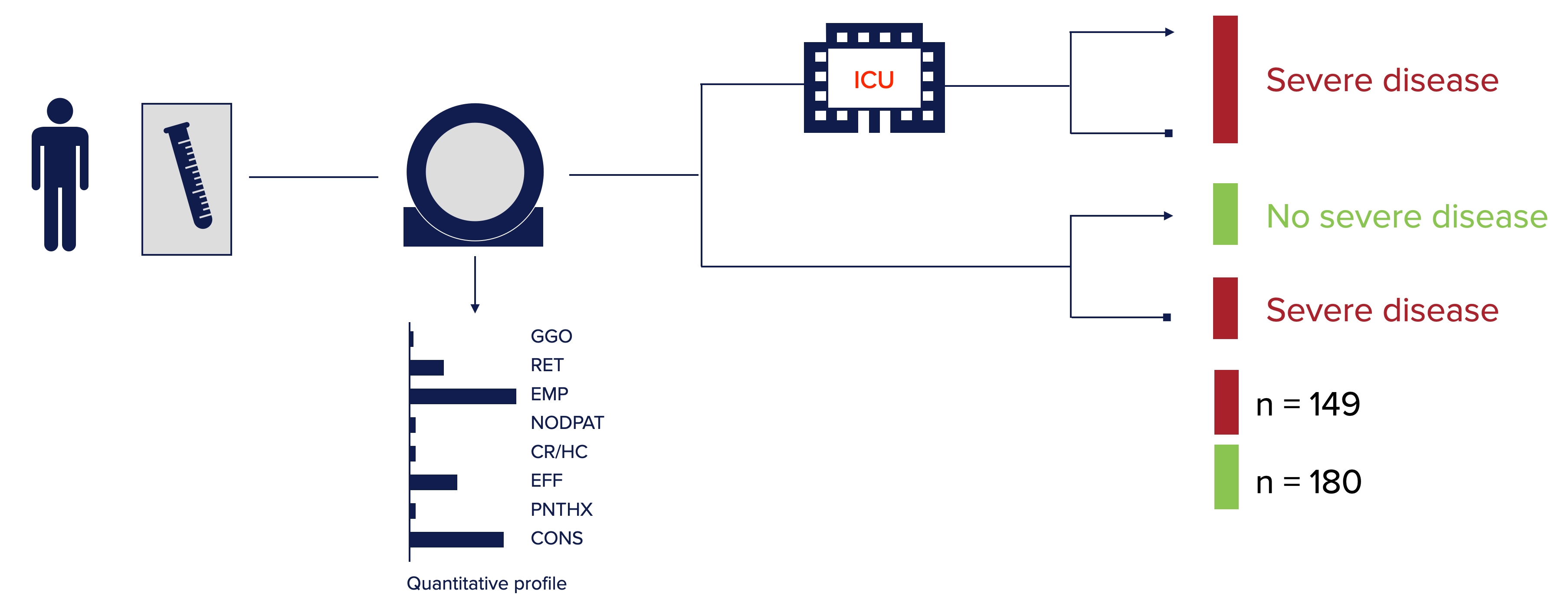
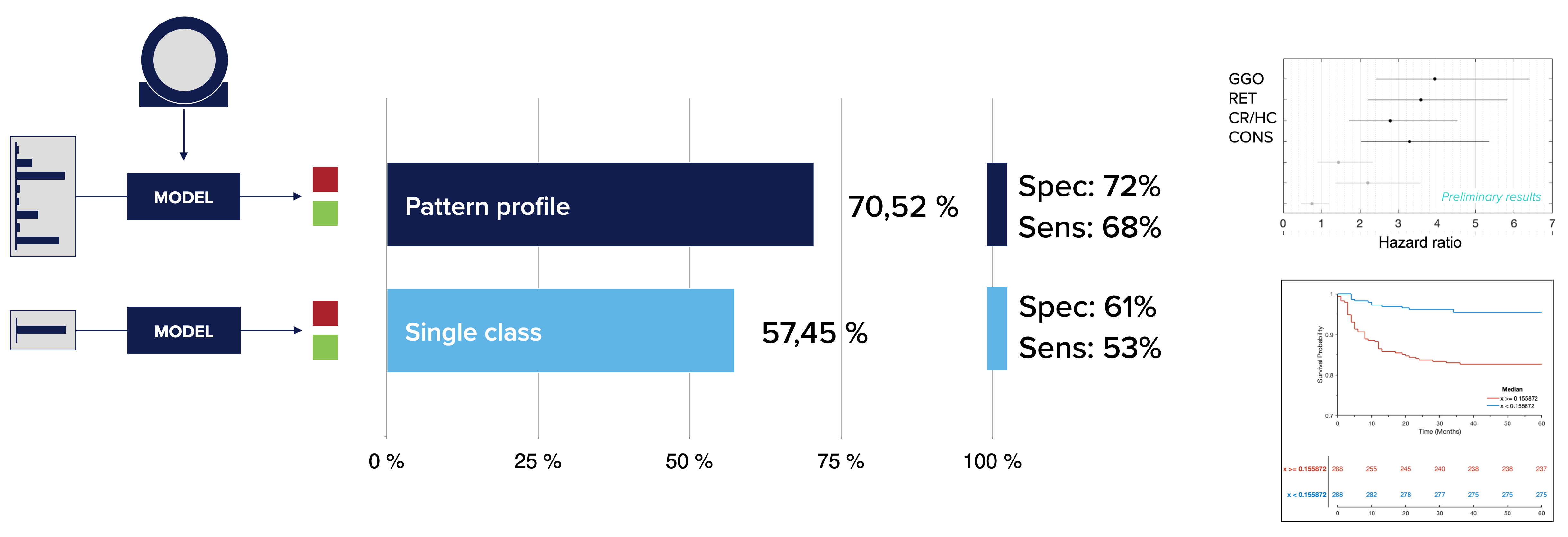
An example of how machine learning can use imaging data to predict individual disease course. Presented at the Annual Scientific Meeting of European Society of Thoracic Imaging 2022 in Oxford, UK by Jeanny Pan et al: Prediction of disease severity in COVID-19 patients identifies predictive disease patterns in lung CT. We could show that quantitative profiles are predictive for future disease course. Importantly, using not only the ratio of normal compared to non-normal tissue but instead the profile of individual disease patterns in the lung improved prediction performance. The study analysed data of 19 hospitals, with overall 329 COVID-19 patients. Based on the quanitative profile obtained from an initial CT scan comprising GGO, reticular pattern, emphysema, nodular pattern, coarse reticulation, effusion, pneumothorax, and consolidation the severity of disease was predicted.
Code
Code and models for automatic lung segmentation in CT data used by many others by now (Hofmanninger et al. 2020): code github
Images: Johannes Hofmanninger, Jeanny Pan, Georg Langs
Relevant publications by project members:
- Mitic, B., Seeböck, P., Straub, J., Prosch, H., Langs, G. (2025). Detection of Emerging Infectious Diseases in Lung CT Based on Spatial Anomaly Patterns. In: Machine Learning in Medical Imaging. MICCAI-MLMI 2024. Lecture Notes in Computer Science, vol 15242. Springer, Cham. https://doi.org/10.1007/978-3-031-73290-4_14 arxiv
- Pan, J., Hofmanninger, J., Nenning, K.H., Prayer, F., Röhrich, S., Sverzellati, N., Poletti, V., Tomassetti, S., Weber, M., Prosch, H. and Langs, G., 2022. Unsupervised machine learning identifies predictive progression markers of IPF. European Radiology
- Pan, J., Hofmanninger, J., Röhrich, S., Prosch, H., and Langs, G., 2022. Prediction of disease severity in COVID-19 patients identifies predictive disease patterns in lung CT. Presentation at Annual Scientific Meeting of European Society of Thoracic Imaging 2022 in Oxford, UK.
- Heidinger, B.H., Kifjak, D., Prayer, F., Beer, L., Milos, R.I., Röhrich, S., Arndt, H. and Prosch, H., 2020. Radiological manifestations of pulmonary diseases in COVID-19. Der Radiologe.
- Revel, M.P., Parkar, A.P., Prosch, H., Silva, M., Sverzellati, N., Gleeson, F. and Brady, A., 2020. COVID-19 patients and the Radiology department–advice from the European Society of Radiology (ESR) and the European Society of Thoracic Imaging (ESTI). European Radiology, 30(9), pp.4903-4909.
- Lang, C., Jaksch, P., Hoda, M.A., Lang, G., Staudinger, T., Tschernko, E., Zapletal, B., Geleff, S., Prosch, H., Gawish, R. and Knapp, S., 2020. Lung transplantation for COVID-19-associated acute respiratory distress syndrome in a PCR-positive patient. The Lancet Respiratory Medicine, 8(10), pp.1057-1060.
- Roberts, M., Driggs, D., Thorpe, M., Gilbey, J., Yeung, M., Ursprung, S., Aviles-Rivero, A.I., Etmann, C., McCague, C., Beer, L. and Weir-McCall, J.R., 2021. Common pitfalls and recommendations for using machine learning to detect and prognosticate for COVID-19 using chest radiographs and CT scans. Nature Machine Intelligence, 3(3), pp.199-217.
- Hofmanninger, J., Prayer, F., Pan, J., Röhrich, S., Prosch, H. and Langs, G., 2020. Automatic lung segmentation in routine imaging is primarily a data diversity problem, not a methodology problem. European Radiology Experimental, 4(1), pp.1-13.
- Röhrich, S., Hofmanninger, J., Negrin, L., Langs, G. and Prosch, H., 2021. Radiomics score predicts acute respiratory distress syndrome based on the initial CT scan after trauma. European Radiology, pp.1-11.
- Röhrich, S., Hofmanninger, J., Prayer, F., Müller, H., Prosch, H. and Langs, G., 2020. Prospects and Challenges of Radiomics by Using Nononcologic Routine Chest CT. Radiology: Cardiothoracic Imaging, 2(4), p.e190190.
Images: MUW/Hofmanninger