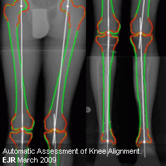
Automatic assessment of the knee alignment angle on full-limb radiographs / Mar 2009

In this study a fully automatic assessment of the knee alignment angles in full-limb radiographs was developed and compared to manual standard of reference measurements in a prospective manner. The data consisted of 28 knees which were gathered from total-leg radiographs of 15 patients (12 males and 3 females with a mean age of 29.4+/-6.9 years) consecutively. For statistical evaluation, a leave-one-out cross-validation was performed. The pattern recognition and consequently the fully automatic assessment were successful in all patients. The automatically measured angles highly correlated with the standard of reference (r=0.989). The mean absolute difference was 0.578 degrees (95% CI: 0.399-0.757 degrees ). 82% of the angles differed less than 1 degrees from the standard of reference, 46% differed less than 0.5 degrees and 31% differed less than 0.2 degrees . The automatic method showed a high agreement between repeated measurements (+0.515 degrees to -0.429 degrees ). The automatic assessment of alignment angles in full-limb radiographs were equal to the manual assessment. No measurement related user interaction was necessary to achieve results.
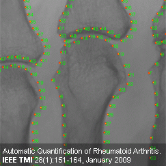
Automatic Quantification of Rheumatoid Arthritis | Jan 2009

Rheumatoid arthritis (RA) is a chronic disease that affects and potentially destroys the joints of the appendicular skeleton. The precise and reproducible quantification of the progression of joint space narrowing and the erosive bone destructions caused by RA is crucial during treatment and in imaging biomarkers in clinical trials. Current manual scoring methods exhibit high inter-reader variability, even after intensive training, and thus, impede the efficient monitoring of the disease.
We propose a fully automatic quantitative assessment of the radiographic changes that result from RA, to increase the accuracy, reproducibility, and speed of image interpretation. Initial joint location estimates are obtained by local linear mappings based on texture features. Bone contours are delineated by active shape models comprised of statistical models of bone shape and local texture. These models are refined by snakes which increase the accuracy and allow for a fitting of pathological deviations from the training population. The method then measures joint space widths and detects erosions on the bone contour. Joint space widths are measured with a coefficient of variation of 2% to 7% for repeated measurements and erosion detection exhibits an area under the ROC curve of 0.89. Model landmarks serve as a reference system along the contour. These landmarks enable the definition of joint regions and more specific follow-up monitoring. The automatic quantification allows for a remote analysis, relevant for multi-center clinical trials, and reduces the workload of clinical experts since parts of the process can be managed by non-expert personnel.
Images:MUW/Langs, Langs
