In our neuroimaging studies on adults, we investigate the functional organisation of the human brain based on functional magnetic resonance imaging (fMRI) in healthy as well as diseased subjects.
Functional mapping to study brain evolution

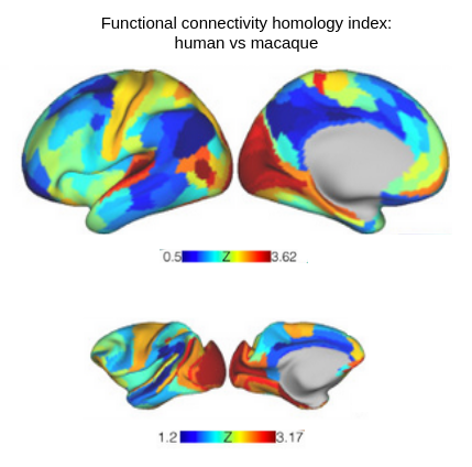
Mapping functional organisation Although functional organisation of the human brain is the same across individuals on the coarse scale, such as the occipital lobe beeing the primary part involved in basic processsing of vision, a substantial amount of inter-subject variability in functional organsiation exists. Mapping functional organisation on the individual level is key in neuroscience research and clincial intervention. We developed several approaches for functional mapping, allowing functional aligment of human subjects or even across species. (Langs et al, 2016, Nenning et al, 2017, Xu and Nenning et al, 2020, best NeuroImage-paper in 2020)

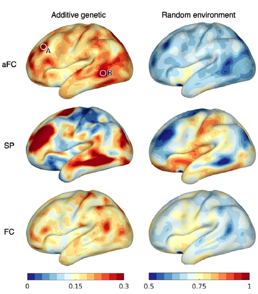
Genetic and environmental influences on variability Based on fMRI data of twins, we investigated the influence of genes and environment on variabilty of fine-grained spatial layout of function, as well as connectivity strength between areas. Functional aligment was used to disentangle function and anatomy. We observed that genetic influence on variability of the spatial layout is inhomogeneous across the cortex and tends to increase with variability itself. Genetic influence on variability of connectivity strength, on the other hand, was more homogeneous across the cortex and lower everywhere than environmental influence. (Burger et al, 2022)
Code for estimating genetic and environmental contribution available: code releases
Machine learning in clinical neuroimaging

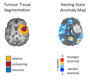
Glioblastoma leads to alterations of the functional connectome Glioblastoma is one of the deadliest cancer types and effects neurocognitive function of patients. Based on longitudinal fMRI data of glioblastoma patients gathered during treatment and disease progression, we observed global impact on the functional connectome. Disruptions are linked to functional proximity rather than anatomical distance to tumour regions. (Nenning et al. 2020)

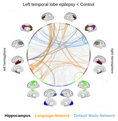
Language connectome in temporal lobe epilepsy Temporal lobe epilepsy may lead to language related impairments, such as difficulties in object naming. In several studies we investigated changes in the functional language connectome induced by epilepsy, or reorganisation after temporal lobe resection. In one study we invesitgated the impact of a diseased left and right hippocampus and its integration with task-positive and task-negative language networks. (Nenning et al, 2021, 2022)
Publications
- K-H Nenning, P Thompson, M Yogarajah, A McEvoy, M Schwarz, K Trimmel, G Kasprian, M Koepp, G Langs, J Duncan, S Bonelli: Functional reorganization of language networks after temporal lobe resection. Epilepsia (2022)
- Bianca Burger, Karl-Heinz Nenning, Ernst Schwartz, Daniel S Margulies, Alexandros Goulas, Hesheng Liu, Simon Neubauer, Justin Dauwels, Daniela Prayer, Georg Langs: Disentangling cortical functional connectivity strength and topography reveals divergent roles of genes and environment. NeuroImage (2022)
- K Nenning, P Thompson, M Yogarajah, A McEvoy, M Schwarz, K Trimmel, G Kasprian, M Koepp, G Langs, J Duncan, S Bonelli-Nauer: Temporal Lobe Resection Reorganizes Language Networks in Patients with Epilepsy. European Journal of Neurology (2021)
- Karl-Heinz Nenning, Olivia Fösleitner, Ernst Schwartz, Michelle Schwarz, Victor Schmidbauer, Gudrun Geisl, Christian Widmann, Susanne Pirker, Christoph Baumgartner, Daniela Prayer, Ekaterina Pataraia, Lisa Bartha-Doering, Georg Langs, Gregor Kasprian, Silvia B Bonelli: The impact of hippocampal impairment on task-positive and task-negative language networks in temporal lobe epilepsy. Clinical Neurophysiology (2021)
- Furtner J., Nenning KH., Roetzer T., Gesperger J., Seebrecht L., Weber M., Grams A., Leber S., Marhold F., Sherif C., Trenkler J., Kiesel B., Widhalm G., Asenbaum U., Woitek R., Berghoff A., Prayer D., Langs G., Preusser M. & Wöhrer A. 2021. Evaluation of the Temporal Muscle Thickness as an Independent Prognostic Biomarker in Patients with Primary Central Nervous System Lymphoma. Cancers, 13(3), p.566.
- Ting Xu, Karl-Heinz Nenning, Ernst Schwartz, Seok-Jun Hong, Joshua T Vogelstein, Alexandros Goulas, Damien A Fair, Charles E Schroeder, Daniel S Margulies, Jonny Smallwood, Michael P Milham, Georg Langs: Cross-species functional alignment reveals evolutionary hierarchy within the connectome. NeuroImage (2020), best NeuroImage-paper in 2020
- Karl-Heinz Nenning, Ting Xu, Ernst Schwartz, Jesus Arroyo, Adelheid Woehrer, Alexandre R Franco, Joshua T Vogelstein, Daniel S Margulies, Hesheng Liu, Jonathan Smallwood, Michael P Milham, Georg Langs: Joint embedding: A scalable alignment to compare individuals in a connectivity space. NeuroImage (2020)
- Karl-Heinz Nenning, Julia Furtner, Barbara Kiesel, Ernst Schwartz, Thomas Roetzer, Nikolaus Fortelny, Christoph Bock, Anna Grisold, Martha Marko, Fritz Leutmezer, Hesheng Liu, Polina Golland, Sophia Stoecklein, Johannes A Hainfellner, Gregor Kasprian, Daniela Prayer, Christine Marosi, Georg Widhalm, Adelheid Woehrer, Georg Langs: Distributed changes of the functional connectome in patients with glioblastoma. Nature Scientific Reports (2020)
- Li M., Wang D.,Ren J., Langs G., Stoecklein S., Brennan B.P., Lu J., Chen H., Liu H. Performing group-level functional image analyses based on homologous functional regions mapped in individuals, PLoS Biology, (2019, in press)
- K.H. Nenning, H. Liuc, S. Ghoshd, M. Sabuncu, E. Schwartz, G. Langs: Diffeomorphic Functional Brain Surface Alignment: Functional Demons. NeuroImage (2017)
- G. Langs, D. Wang, P. Golland, S. Mueller, R. Pan, M. Sabuncu, W. Sun, K. Li, H. Liu: Identifying Shared Brain Networks in Individuals by Decoupling Functional and Anatomical Variability. Cerebral Cortex (2016)
- D. Wang, R. L. Buckner, M. D. Fox, D. J. Holt, A. J. Holmes, S. Stoecklein, G. Langs, R. Pan, T. Qian, K. Li, J. T. Baker, S. M. Stufflebeam, K. Wang, X. Wang, B. Hong, and H. Liu: Parcellating cortical functional networks in individuals. Nature Neuroscience (2015)
- Karl-Heinz Nenning, Kathrin Kollndorfer, Veronika Schöpf, Daniela Prayer, and Georg Langs: Multi-Subject Manifold Alignment of Functional Network Structures via Joint Diagonalization. Advances in Information Processing in Medical Imaging, IPMI 2015
Images: MUW/Nenning, Burger, Nenning, Nenning
